
Back & Spine Pain
Understanding Back Pain —
Finding the Source
Persistent back pain is rarely just about your back. ROOT METHOD traces every symptom — localised pain, radiating sciatica, morning stiffness, or neurological changes — to its structural, neurological, or mechanical origin.
Clinical Overview
What is Back Pain?
Back pain is one of the most common clinical presentations worldwide — but it is a symptom, not a diagnosis. The term covers a wide spectrum of experiences, from acute muscle strain that resolves in days to complex chronic pain that has persisted for years without a clear explanation.
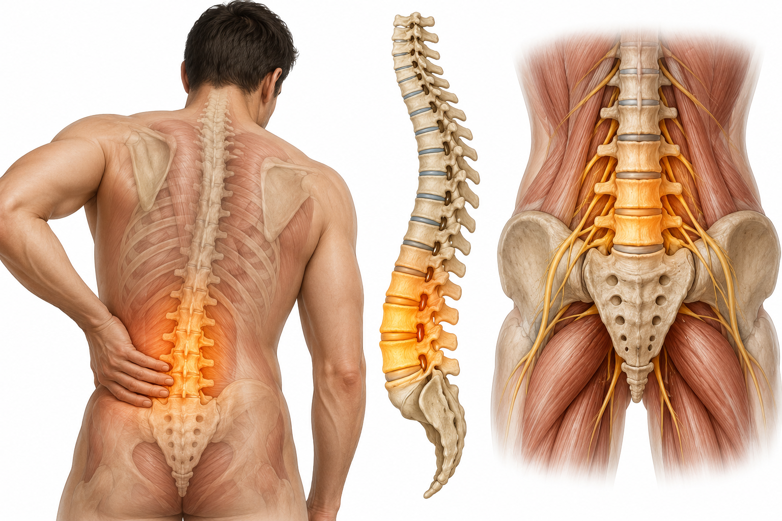
The spine is a highly integrated structure: vertebrae, intervertebral discs, facet joints, ligaments, muscles, and a network of nerve roots all working in coordination. When any component is under stress — from degeneration, compression, inflammation, or mechanical loading — pain can arise. Crucially, the location of pain does not always correspond to the location of its source.
Acute back pain typically follows a specific event — a fall, a sudden movement, or a period of sustained physical load. Chronic back pain persists beyond 12 weeks and is more often the product of underlying structural or neurological issues that have not been identified and addressed. It is chronic back pain — particularly pain that has not responded to standard treatment — that ROOT METHOD is best positioned to assess.
Clinical Presentation
Common Symptoms of Back Pain
Back pain rarely presents as a single, isolated sensation. Most patients experience a combination of the following — each of which carries diagnostic significance.
Localised Pain
Sharp, dull, aching, or burning pain confined to a specific region of the back — commonly the lower lumbar area, but also the thoracic spine or sacroiliac joint. The character and location of localised pain help identify the affected structure.
Radiating Pain
Pain that travels from the back into the buttock, thigh, calf, or foot — commonly referred to as sciatica. Radiating pain typically indicates nerve root irritation or compression at a specific spinal level, and its distribution often points directly to the source.
Morning Stiffness
Significant stiffness upon waking that eases within 30 to 60 minutes of movement. Morning stiffness is associated with inflammatory conditions and facet joint dysfunction. Stiffness that takes longer than an hour to improve warrants specific clinical attention.
Muscle Spasm
Involuntary, sustained contraction of the paraspinal or gluteal muscles that causes acute pain and restriction of movement. Muscle spasm is typically a protective response to an underlying structural problem — treating the spasm without addressing its cause produces only temporary relief.
Neurological Symptoms
Numbness, tingling, pins and needles, or weakness in one or both legs. These symptoms arise when nerve tissue is under compression or irritation. Their specific distribution — which dermatome or myotome is affected — identifies the spinal level responsible.
Referred Pain
Pain felt in the hip, groin, abdomen, or pelvis that originates in the lumbar spine or sacroiliac joint. Referred pain frequently misleads both patients and clinicians — it is often investigated in the wrong location before the spinal origin is recognised.
Clinical Aetiology
Common Causes of Back Pain
Identifying the correct cause is the central clinical challenge of back pain management. Many patients carry multiple contributing factors — which is why a systematic investigation is essential.
Disc Pathology
Intervertebral disc herniation, bulge, or degeneration is one of the most common structural causes of back and leg pain. A compromised disc can compress adjacent nerve roots — producing characteristic radiating pain patterns that guide spinal level identification.
Nerve Root Compression
Radiculopathy occurs when a spinal nerve root is compressed or chemically irritated — most commonly by a herniated disc, bone spur, or narrowed foramen. The resulting pain, numbness, and weakness follow a predictable dermatomal pattern that is diagnostically precise when properly mapped.
Facet Joint Degeneration
The facet joints connect adjacent vertebrae and guide spinal movement. Arthritic degeneration of these joints produces a characteristic deep, aching pain that is often worse with extension and rotation. Facet-mediated pain is commonly misdiagnosed or attributed entirely to disc pathology.
Inflammatory Conditions
Ankylosing spondylitis, inflammatory arthritis, and other spondyloarthropathies cause back pain with a distinctive pattern: insidious onset in younger patients, significant morning stiffness lasting more than an hour, and improvement with activity rather than rest. These conditions require specific clinical recognition.
Sacroiliac Joint Dysfunction
The sacroiliac joint connects the sacrum to the pelvis and is a frequently overlooked source of low back and buttock pain. Dysfunction here produces pain that is often mistaken for lumbar disc pathology — it typically presents below L5, radiates into the buttock, and is aggravated by single-leg loading activities.
Postural and Mechanical Dysfunction
Sustained postural load, occupational postures, muscular imbalance, and altered movement patterns place disproportionate mechanical stress on spinal structures. Over time, this creates micro-damage and compensatory dysfunction — pain that builds gradually and has no single identifiable onset event.
Clinical Guidance
When to Seek Professional Assessment
Not all back pain requires specialist input — but some does, and some requires it urgently. Knowing the difference matters.
Book a Clinical Assessment If…
- Your back pain has persisted for more than 6 weeks without meaningful improvement
- Pain recurs regularly despite rest, physiotherapy, or medication
- You experience radiating pain into the buttock, leg, or foot
- Stiffness significantly limits your daily activity, sleep, or work performance
- You have received multiple diagnoses without a clear clinical explanation
- Your pain began after an injury and has not fully resolved within expected timeframes
Seek Emergency Care Immediately If…
- You experience sudden loss of bladder or bowel control — this may indicate cauda equina syndrome, a medical emergency
- You develop progressive weakness or numbness in both legs simultaneously
- Back pain follows significant trauma such as a fall, road accident, or direct impact
- You have unexplained weight loss alongside new-onset back pain
- Pain is severe, unrelenting, and worsens at night when lying down
The ROOT METHOD Difference
How ROOT METHOD Approaches Back Pain
The most critical clinical question in back pain is not "how bad is it?" — it is "what is causing it?" A disc herniation, a facet joint, a sacroiliac joint, an inflamed nerve root, and a postural compensation pattern all produce pain that patients describe similarly. They require completely different treatments.
ROOT METHOD approaches each patient's back pain as a clinical investigation — not a treatment pathway. Before any intervention is considered, the assessment works to answer:
- Which spinal structure is the primary pain generator — disc, facet, nerve root, or sacroiliac joint?
- Is there neurological involvement, and if so, at which spinal level?
- Is the pain mechanical, inflammatory, referred, or a combination?
- What postural, occupational, or biomechanical factors are sustaining the problem?
- Are there adjacent structures — hip, pelvis, kidney — contributing to the presentation?
Your First Visit
What to Expect During Your Back Pain Assessment
A ROOT METHOD back pain assessment is a 90-minute clinical appointment. Here is what happens at each stage.
-
Before You Arrive
Pre-Visit Intake Form
You will complete the ROOT METHOD 8-step intake form before your appointment. This covers your full pain history, onset and duration, prior treatments, medication, and how your symptoms vary with posture and activity. Reviewing this in advance allows Dr. Umer to begin clinical reasoning before you enter the room.
-
Stage 1
Postural and Movement Assessment
Dr. Umer observes your posture, spinal alignment, and movement patterns in standing and during specific motions — flexion, extension, lateral flexion, and rotation. Compensatory movement strategies and range-of-motion restrictions are documented. The position that most reliably reproduces your pain is identified.
-
Stage 2
Neurological and Structural Examination
A systematic neurological examination tests sensation, reflexes, and muscle strength in the lower limbs. Nerve tension tests — including straight leg raise and femoral nerve stretch — assess nerve root involvement. Palpation of the spine, sacroiliac joint, and surrounding musculature identifies localised tenderness and tissue changes.
-
Stage 3
Diagnosis and Protocol Discussion
Dr. Umer reviews all findings with you and explains the clinical reasoning in plain language. You receive a specific diagnosis — or a differential diagnosis where multiple contributors are present — and a personalised treatment protocol. You leave the appointment knowing what is causing your pain and what the plan is to address it at source.

Common Questions
Questions About Back Pain and ROOT METHOD
What is the most common cause of lower back pain?
There is no single answer — which is precisely the clinical problem. Disc herniation, facet joint degeneration, sacroiliac joint dysfunction, and muscular imbalance are all common, and they frequently coexist in the same patient. In clinical practice, the most common mistake is attributing all lower back pain to disc pathology because an MRI shows a disc herniation — without determining whether that disc is actually causing the patient's specific symptom pattern.
ROOT METHOD's approach is to identify which structure is the primary pain generator in your individual case — not to default to the most common diagnosis.
Do I need an MRI before my assessment?
No — and in many cases, having an MRI before a thorough clinical examination can be misleading. MRI findings, including disc herniations and degenerative changes, are extremely common in people without back pain and do not, by themselves, constitute a diagnosis. Imaging findings must be interpreted in the context of a clinical examination to determine whether they are actually responsible for your symptoms.
If you have prior MRI reports, please bring them. Dr. Umer will review them as part of the assessment. If imaging is genuinely needed after the clinical examination, it will be requested with a specific clinical question attached.
What is sciatica, and how does ROOT METHOD address it?
Sciatica is pain that travels from the lower back or buttock into the leg — following the distribution of the sciatic nerve. It is a symptom, not a diagnosis. The underlying cause may be a disc herniation compressing the L4, L5, or S1 nerve root; spinal stenosis narrowing the neural canal; a piriformis syndrome compressing the sciatic nerve in the buttock; or a sacroiliac joint referral pattern that mimics nerve pain.
ROOT METHOD distinguishes between these causes through clinical examination and nerve tension testing. Treatment depends entirely on the identified origin — which is why the same sciatica can require completely different management in different patients.
I have had physiotherapy and injections with no lasting improvement. Can ROOT METHOD help?
This is one of the most common presentations at ROOT METHOD. Physiotherapy and injections are effective when applied to the correct diagnosis — but when the root cause has not been identified, even the best technical execution of a treatment protocol will not produce lasting results. The intervention was not wrong; the target was.
ROOT METHOD begins by examining what the prior treatments were targeting and whether the clinical evidence supports that target. In many cases, a different pain generator is identified — one that has not previously been investigated — and a revised protocol can produce significant improvement.
How long does back pain take to resolve with the right treatment?
Resolution time depends on the underlying cause, duration of the problem, and the patient's response to treatment. Acute back pain with a clear mechanical cause often improves significantly within 4 to 8 weeks of appropriately targeted treatment. Chronic back pain that has persisted for months or years typically requires a longer course — but most patients with an accurate diagnosis begin to notice measurable improvement within the first 2 to 4 weeks.
Clear clinical benchmarks are established at the outset of every ROOT METHOD protocol. Progress is measured against these benchmarks, and protocols are adjusted if the expected response is not occurring.
What should I avoid doing while waiting for my assessment?
Avoid complete bed rest — prolonged inactivity is well-documented to worsen back pain outcomes. Gentle movement, within pain tolerance, is generally beneficial. Avoid activities that significantly increase your pain, particularly sustained flexion postures (prolonged sitting or bending) if these are your main aggravating factors.
Continue any medication your GP has prescribed. If you are awaiting an urgent assessment for neurological symptoms — leg weakness, numbness, or bladder changes — seek advice from your GP or emergency services promptly rather than waiting for a scheduled appointment.
Back Pain Has a Source. We Find It.
Most patients with persistent back pain have never received a complete clinical investigation of what is driving it. ROOT METHOD identifies the origin of your pain — and builds a treatment plan that addresses it at source, not at the symptom.