
Knee Pain
Understanding Knee Pain —
Finding the Source
The knee is the largest joint in the body and among the most mechanically loaded. Pain can arise from multiple structures within and around the joint — and misidentifying the source leads to treatments that relieve symptoms temporarily without addressing the underlying problem.
Clinical Overview
What is Knee Pain?
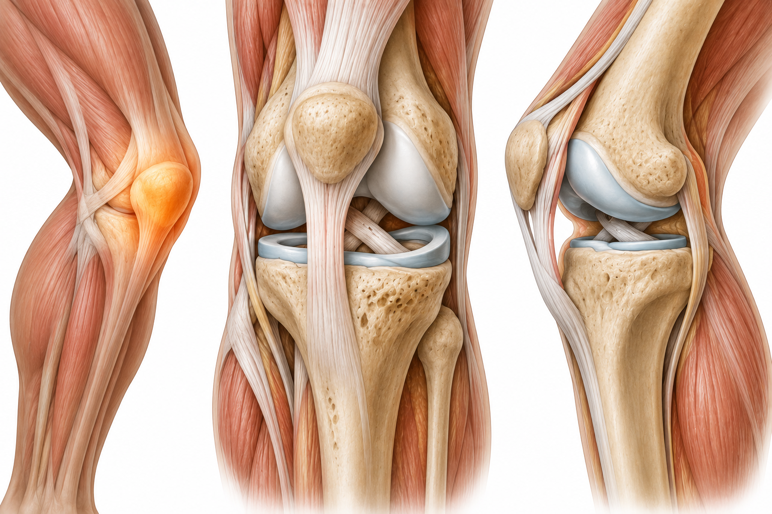
The knee joint comprises the tibiofemoral and patellofemoral articulations, supported by the anterior and posterior cruciate ligaments, medial and lateral collateral ligaments, the medial and lateral menisci, and an extensive surrounding musculature. Pain can originate from any of these structures — cartilage, menisci, ligaments, tendons, bursa, or the patellofemoral joint — each producing a characteristic clinical pattern when examined systematically.
The location of pain within the knee is a primary diagnostic clue. Anterior knee pain — especially with stair use — points toward the patellofemoral joint or patellar tendon. Medial joint line pain suggests medial meniscus or medial collateral ligament. Lateral pain may indicate the lateral meniscus, IT band, or lateral compartment cartilage. Generalised joint swelling following activity suggests an intra-articular pathology requiring careful structural assessment.
An important principle: the knee rarely fails in isolation. Hip abductor weakness, foot pronation, and altered movement mechanics contribute to excessive knee loading and drive many common knee conditions. A complete knee assessment must account for the entire lower limb kinetic chain, not only the structures within the joint itself.
Clinical Presentation
Common Symptoms of Knee Pain
The location, behaviour, and pattern of knee pain each provide critical structural diagnostic information when interpreted by a trained clinician.
Anterior Knee Pain
Pain at the front of the knee — around or behind the patella — that is classically worsened by stair climbing, squatting, kneeling, and prolonged sitting (cinema sign). This anterior pattern strongly suggests patellofemoral joint dysfunction or patellar tendinopathy, and distinguishes itself from posterior or joint-line pain.
Medial or Lateral Knee Pain
Joint-line pain on the inner or outer aspect of the knee is characteristic of meniscal pathology — acute tear or degenerative tear. Medial collateral ligament sprain produces medial pain with valgus stress. Lateral pain with activity may indicate the lateral meniscus, IT band syndrome, or lateral compartment cartilage disease.
Knee Swelling and Joint Effusion
Visible or palpable swelling within the knee joint indicates an intra-articular process. A rapid haemarthrosis (blood in the joint) following trauma strongly suggests ligament rupture or osteochondral fracture. Gradual inflammatory effusion is characteristic of meniscal tear, osteoarthritis flare, or crystal arthropathy, and requires clinical distinction.
Locking or Giving Way
True mechanical locking — inability to fully extend the knee — indicates a displaced meniscal fragment or loose body blocking joint movement. Giving way (the knee buckling) may indicate ligamentous instability (ACL insufficiency) or quadriceps inhibition. Both symptoms require systematic assessment and should not be attributed to weakness or fatigue without structural investigation.
Pain After Prolonged Sitting Then Standing
Stiffness and pain in the knee after sustained sitting — the cinema sign — that eases after a few steps. This pattern is highly characteristic of patellofemoral joint dysfunction and reflects increased patellofemoral joint pressure during prolonged flexion. It is one of the most reliable indicators of anterior knee pathology in clinical practice.
Deep Aching Pain at Rest
Persistent, deep aching in the knee that is present at rest and worsens after activity — characteristic of osteoarthritis, particularly involving the medial tibiofemoral compartment. Night pain that disrupts sleep in OA indicates a more severe or inflammatory pattern, and distinguishes arthritic pain from mechanical pain that typically resolves with rest.
Clinical Aetiology
Common Causes of Knee Pain
Knee pain arises from distinct structural sources — each with a characteristic clinical signature that guides accurate diagnosis and appropriate treatment.
Patellofemoral Pain Syndrome
The most common cause of anterior knee pain in young active adults — arising from abnormal tracking of the patella over the femoral groove. Driven by hip abductor weakness, quadriceps imbalance, foot pronation, and training load errors. Produces characteristic anterior knee pain with stair use, squatting, and prolonged sitting. Frequently mismanaged with rest alone without addressing the biomechanical drivers.
Meniscal Tear
The menisci act as shock absorbers and load distributors within the tibiofemoral joint. Acute tears occur from twisting injuries under load; degenerative tears develop with age in the absence of a specific event. Produces medial or lateral joint-line pain, joint effusion, and sometimes locking or giving way. McMurray's and Thessaly tests aid clinical confirmation.
Osteoarthritis
Degenerative loss of articular cartilage — most commonly in the medial tibiofemoral compartment — producing progressive knee pain, morning stiffness easing within 30 minutes, crepitus, and restriction of flexion. The medial compartment bears greater load due to normal varus alignment, making it disproportionately affected. OA severity on imaging does not reliably predict functional limitation.
Patellar Tendinopathy
Degenerative change within the patellar tendon — most commonly at the inferior pole of the patella — producing anterior knee pain that is specifically reproduced by loading in knee flexion (landing, jumping, squatting). Common in jumping sports (volleyball, basketball) but also affects those with sudden increases in activity or stair-climbing demands. Clinical diagnosis confirmed by Victoriano test and arc of pain assessment.
ACL or Ligament Injury
Anterior cruciate ligament (ACL) rupture typically occurs during a non-contact deceleration, pivoting, or landing mechanism — producing immediate haemarthrosis, instability, and giving way. Posterior cruciate, medial collateral, and lateral collateral ligament injuries each have specific mechanisms and clinical test patterns. Grading and specific ligament identification determines the rehabilitation pathway and surgical decision.
Pes Anserine Bursopathy
Inflammation of the pes anserine bursa on the medial aspect of the proximal tibia, producing medial knee pain just below the joint line — often confused with meniscal or OA pain. Common in overweight patients, those with knee OA, and swimmers (due to the breaststroke kick). Characteristically worsened by climbing stairs and tender on direct palpation below the medial joint line.
Clinical Guidance
When to Seek Professional Assessment
Knee pain that limits activity, is accompanied by swelling or instability, or fails to resolve within expected timeframes requires clinical investigation to identify the structural source.
Book a Clinical Assessment If…
- Knee pain has persisted for more than 6 weeks despite rest and activity modification
- You experience knee swelling — even mild or intermittent — without clear explanation
- The knee gives way, locks, or catches during normal activity
- Pain significantly limits stair climbing, squatting, kneeling, or sport participation
- You have received a diagnosis of knee pain without a specific structural explanation
- Knee pain recurs after each return to activity, suggesting an unaddressed underlying cause
Seek Emergency Care Immediately If…
- Acute haemarthrosis (rapid, tense swelling) after trauma — possible ACL rupture, osteochondral fracture, or peripheral meniscal tear requiring prompt assessment
- True mechanical locking — inability to extend the knee — may indicate a displaced meniscal fragment requiring urgent orthopaedic assessment
- Hot, erythematous, acutely swollen knee with fever — possible septic arthritis or crystal arthropathy requiring immediate evaluation
- Knee injury with gross deformity or inability to weight-bear — possible fracture or dislocation
- Popliteal artery pulse absent after knee dislocation — vascular emergency

The ROOT METHOD Difference
How ROOT METHOD Approaches Knee Pain
Every knee assessment at ROOT METHOD begins with a full lower limb kinetic chain evaluation — because knee pain is rarely caused by the knee alone. Hip abductor strength, patellar tracking, quadriceps-to-hamstring balance, and foot mechanics each contribute to the forces transmitted through the knee with every step.
The structural assessment applies a targeted sequence of clinical tests to identify the specific pain-generating structure before any treatment recommendation is made:
- McMurray test and Thessaly test — assessing medial and lateral meniscal integrity
- Lachman and anterior drawer tests — evaluating ACL integrity and grading insufficiency
- Patellar grind, tilt, and apprehension tests — assessing patellofemoral joint mechanics and pain
- Joint-line palpation — localising medial, lateral, and posterior joint tenderness
- Quadriceps and VMO assessment — identifying medial muscle weakness contributing to patellofemoral maltracking
Your First Visit
What to Expect During Your Knee Pain Assessment
A ROOT METHOD knee assessment is a 90-minute clinical appointment identifying the specific structural source of your knee pain and its contributing biomechanical factors.
-
Before You Arrive
Pre-Visit Intake Form
You complete the ROOT METHOD intake form covering your knee pain location, onset, mechanism of injury if applicable, aggravating and easing factors, prior imaging and diagnoses, and your activity level. The precise location of pain within the knee — anterior, medial, lateral, or diffuse — begins structuring the clinical reasoning before your appointment.
-
Stage 1
Neurological Examination
Lumbar neurological screening excludes L3, L4 radiculopathy as a contributing source of knee pain — a common but frequently missed differential. Lower limb sensation, reflexes, and myotomal strength testing are performed systematically before knee-specific tests begin.
-
Stage 2
Structural Examination
A systematic sequence of ligament, meniscal, patellofemoral, and palpation tests identifies the structural fault. Lower limb biomechanics — hip strength, patellar tracking, gait pattern — are assessed as part of the same session. Swelling, crepitus, and range-of-motion restriction are documented and graded.
-
Stage 3
Diagnosis & Protocol
Dr. Umer reviews findings and provides a specific structural diagnosis with a personalised treatment plan — including imaging requests where indicated, load management guidance, rehabilitation targets, and clear return-to-activity criteria. You leave the appointment with a clear clinical explanation, not a generic diagnosis of 'knee pain'.

Common Questions
Questions About Knee Pain and ROOT METHOD
How do I know if I have a meniscal tear?
The classic features of a meniscal tear are joint-line pain (medial or lateral), joint effusion appearing within 24–48 hours of injury, and specific provocation by deep knee flexion and rotation. A history of a twisting or loading mechanism under bodyweight, followed by immediate joint-line pain and swelling, is characteristic of an acute meniscal injury. Locking — inability to fully extend the knee — indicates a displaced bucket-handle tear that has folded into the joint.
Clinical tests — McMurray's, Thessaly, and joint-line palpation — are the primary diagnostic tools. MRI is the confirmatory investigation of choice and is requested with the specific clinical question framed by the examination findings. Not all meniscal tears on MRI are symptomatic — particularly in older patients, degenerative meniscal changes are ubiquitous and must be clinically correlated to the patient's specific symptoms before being attributed as the pain source.
What is patellofemoral pain syndrome?
Patellofemoral pain syndrome (PFPS) — sometimes called runner's knee — is the most common cause of anterior knee pain in active people. It arises from abnormal mechanics of the patella tracking through the femoral groove during knee flexion and extension. When the patella tracks laterally — due to hip abductor weakness, quadriceps imbalance (particularly weak VMO), tight lateral soft tissues, or foot pronation — the patellofemoral joint contact forces are distributed unevenly, producing anterior knee pain with activity.
The diagnosis is clinical — there is no specific scan finding that confirms PFPS. The characteristic pattern is anterior knee pain worsened by stair use, squatting, kneeling, and prolonged sitting. Pain is reproduced by patellar grind and compression tests. The treatment approach is fundamentally biomechanical — addressing the proximal and distal drivers of patellar maltracking, not simply resting the knee. This is why PFPS responds poorly to rest alone and requires targeted rehabilitation directed at the identified driving factors.
Does knee OA always lead to surgery?
No — the majority of knee osteoarthritis is managed successfully without surgery, often indefinitely. Conservative management that is appropriately structured — targeted quadriceps and hip strengthening, load management, weight optimisation, and activity modification — consistently reduces pain and improves function in knee OA. Evidence shows that structured exercise is as effective as anti-inflammatory medication for OA pain in many patients, with superior long-term outcomes and no side effects.
Total knee replacement is considered when conservative management has been optimised and fails to provide acceptable pain relief and function. It is a highly effective intervention — but it is not the inevitable destination for every patient with knee OA. The decision to pursue surgical referral is made based on symptom severity, functional limitation, degree of structural destruction, and the patient's response to an adequately delivered conservative programme. ROOT METHOD's role is to ensure that conservative management has been correctly targeted before surgical options are discussed.
Can strengthening exercises reverse cartilage loss?
Cartilage is avascular and has limited intrinsic healing capacity — established cartilage loss does not regenerate through exercise. However, this is not the reason to exercise. Strengthening the muscles surrounding the knee — particularly the quadriceps, gluteals, and hip abductors — reduces the load transmitted through the damaged cartilage, improves joint stability, and reduces pain. This is well-established in clinical research: patients with knee OA who engage in regular targeted strengthening experience significantly less pain than those who are sedentary, even with identical imaging changes.
Exercise also provides broader benefits — improved cardiovascular health, weight management, reduced inflammation — that indirectly affect OA progression and pain levels. The type and intensity of exercise matters: low-impact, high-repetition strengthening with appropriate load progression is more effective and safer than high-impact activity. ROOT METHOD prescribes specific exercise protocols based on the patient's structural diagnosis and current load tolerance, rather than generic exercise advice.
Why does my knee hurt going downstairs?
Descending stairs produces significantly greater patellofemoral joint compressive force than ascending stairs or walking on flat ground — up to 3.5 times body weight. For this reason, pain specifically provoked by going downstairs (more than going up) is a cardinal feature of patellofemoral joint pathology, including both PFPS and patellofemoral osteoarthritis. The eccentric contraction of the quadriceps required to lower the body against gravity loads the patella into the femoral groove with high force.
Descending stairs also loads the medial knee compartment eccentrically, which can reproduce medial OA or meniscal pain — though these are typically distinguished by their location (medial joint line vs anterior). If stair descent specifically reproduces your knee pain, the patellofemoral joint is the most likely primary source, and the assessment is directed accordingly.
Is knee pain related to hip or foot alignment?
Yes — the knee is mechanically linked to both the hip above and the foot below, and dysfunction in either direction can drive knee pain. Hip abductor weakness allows the femur to adduct excessively during single-leg activities — increasing lateral patellofemoral loading and valgus stress on the medial knee. Research consistently shows that strengthening the hip abductors reduces patellofemoral pain, even without directly treating the knee itself.
From below, excessive foot pronation increases tibial internal rotation during walking and running, which increases valgus stress at the knee and alters patellofemoral tracking. Custom orthotics or footwear modification can reduce this tibial torque. ROOT METHOD's assessment includes both hip strength assessment and lower limb alignment analysis — evaluating the Q-angle, gait mechanics, and foot posture — to identify which external factors are driving the knee problem. Treatment that addresses only the knee without these contributing factors is a common reason for incomplete or temporary recovery.
Knee Pain Has a Source. We Find It.
The knee contains many structures — each a potential pain source with its own clinical signature. ROOT METHOD identifies the exact structure at fault and addresses the biomechanical factors that allowed it to fail, preventing the cycle of partial treatment and recurrence.