
Carpal Tunnel Syndrome
Carpal Tunnel Syndrome —
Finding the Source
Carpal tunnel syndrome is the most common peripheral nerve entrapment — yet it is frequently misdiagnosed, over-diagnosed, or missed entirely because the clinical history is not taken with sufficient precision.
Clinical Overview
What is Carpal Tunnel Syndrome?
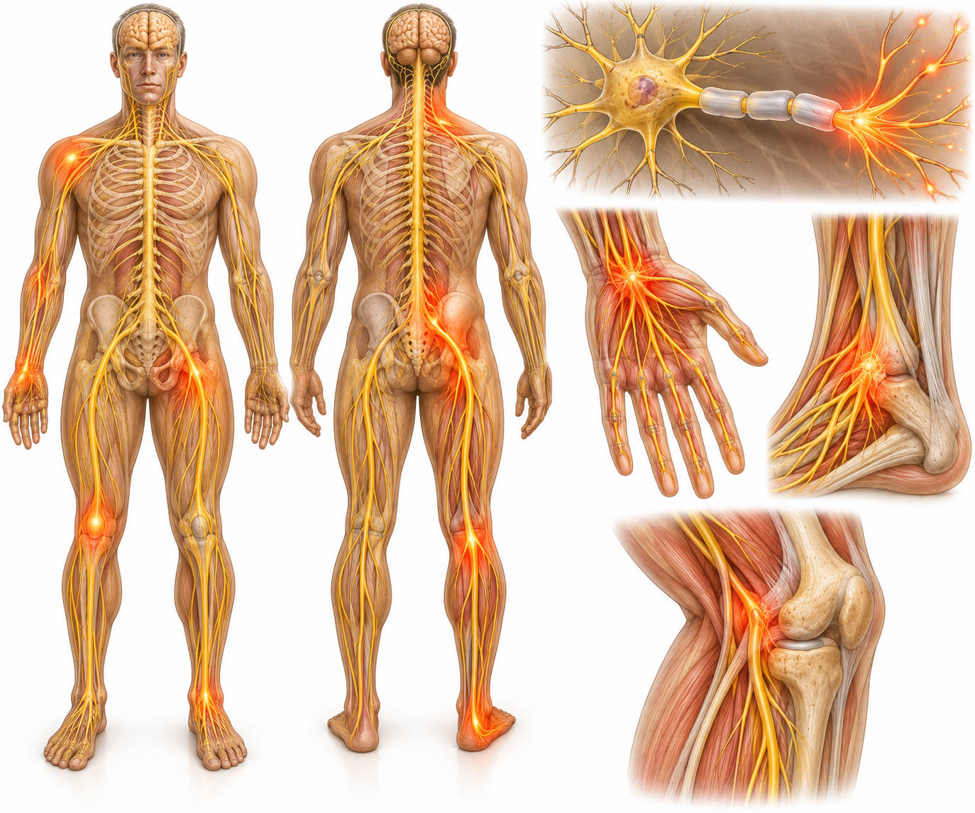
Carpal tunnel syndrome (CTS) occurs when the median nerve is compressed within the carpal tunnel — a narrow channel on the palmar side of the wrist bounded by carpal bones and the transverse carpal ligament. The median nerve supplies sensation to the thumb, index, middle, and radial half of the ring finger, and motor function to the thenar muscles at the base of the thumb. Compression produces a characteristic pattern of symptoms — but the cervical spine, thoracic outlet, and other sites of median nerve compression must be systematically excluded before a diagnosis of CTS is confirmed.
The key diagnostic challenge with CTS is that the median nerve can be compressed at multiple points along its course — from the C6 nerve root in the cervical spine, through the thoracic outlet, to the pronator teres in the forearm, and finally at the wrist. This concept — the double crush phenomenon — means that a patient can have genuine CTS and a contributing cervical radiculopathy simultaneously, and treating only the wrist will produce incomplete recovery.
Clinical diagnosis of CTS is based on a characteristic symptom pattern confirmed by physical examination. Nerve conduction studies provide objective confirmation and grade severity, but should be interpreted alongside the clinical picture rather than as a standalone investigation. A thorough clinical examination — including cervical spine assessment — is the essential first step.
Clinical Presentation
Common Symptoms of Carpal Tunnel Syndrome
The symptom pattern of CTS is characteristic — but must be carefully distinguished from cervical radiculopathy and other peripheral nerve compressions.
Numbness & Tingling in Thumb, Index, and Middle Fingers
The hallmark symptom of CTS — abnormal sensation in the median nerve territory. The ring finger may be partially affected on the radial half. The little finger is spared entirely, which distinguishes CTS from ulnar nerve compression at the elbow. The distribution is highly specific when examined carefully.
Nocturnal Symptoms — Waking with Hand Symptoms
Symptoms typically worsen at night, waking patients from sleep with hand numbness or burning. This nocturnal pattern reflects the sustained wrist flexion that occurs during sleep, increasing pressure within the carpal tunnel and intensifying median nerve compression. Night symptoms are strongly characteristic of CTS.
Symptom Relief by Shaking the Hand
A highly characteristic behaviour — patients instinctively shake or flick the wrist and hand upon waking to relieve symptoms. This flick sign has strong positive predictive value for CTS and represents the patient's attempt to restore blood flow and reduce intraneural pressure through movement.
Weakness of Grip and Pinch (Advanced Cases)
As CTS progresses, weakness of the thenar muscles — abductor pollicis brevis in particular — reduces the ability to oppose the thumb and perform precision pinch tasks. Thenar wasting (visible flattening of the thenar eminence) indicates significant, prolonged compression and the need for prompt clinical action.
Dropping Objects
A consequence of reduced grip strength and altered sensory feedback from the median nerve territory. Patients report unexpectedly losing grip on cups, tools, or phones. Dropping objects is a functional marker of significant CTS severity and typically indicates the need for more active management than conservative measures alone.
Symptoms Worsened by Sustained Wrist Flexion
Activities that maintain the wrist in a flexed position — driving, typing, holding a phone or book — typically reproduce or worsen symptoms. This is the basis of the Phalen's test, which reproduces symptoms by maintaining full wrist flexion for 60 seconds, and confirms that wrist position is mechanically loading the median nerve.
Clinical Aetiology
Common Causes of Carpal Tunnel Syndrome
CTS results from any factor that reduces the space within the carpal tunnel, increases the volume of its contents, or sensitises the median nerve to compression.
Repetitive Wrist Use and Sustained Positions
Prolonged keyboard use, mouse operation, assembly-line work, and any occupation requiring sustained wrist flexion or repetitive grip are among the most common contributing factors. These activities increase pressure within the carpal tunnel and over time sensitise the median nerve to compression at this site.
Fluid Retention
Pregnancy and hypothyroidism both cause fluid retention that increases the volume of the carpal tunnel contents, compressing the median nerve. CTS is extremely common in the third trimester of pregnancy and often resolves postpartum. In hypothyroidism, myxoedematous tissue infiltration directly narrows the available space for the median nerve.
Wrist Arthritis or Prior Fracture
Previous distal radius fracture (Colles' fracture) can narrow the carpal tunnel through malunion, callus formation, or post-traumatic swelling. Osteoarthritis at the wrist, particularly carpometacarpal joint arthritis, produces synovial thickening and osteophytes that reduce tunnel volume and compress the median nerve.
Diabetes Mellitus
Diabetes is a significant risk factor for CTS through two mechanisms: peripheral neuropathy makes nerves more susceptible to compression injury (double crush), and advanced glycation end-products thicken the transverse carpal ligament and surrounding tissues. Diabetic patients with hand symptoms require careful assessment to distinguish CTS from peripheral diabetic neuropathy.
Inflammatory Arthritis
Rheumatoid arthritis, psoriatic arthritis, and other inflammatory conditions produce synovial proliferation within the carpal tunnel — directly compressing the median nerve. CTS may be the presenting feature of an undiagnosed inflammatory arthritis. Systemic inflammatory markers and joint pattern should always be assessed in bilateral or early-onset CTS presentations.
Idiopathic
The majority of CTS cases have no single identifiable cause — they are classified as idiopathic, arising from the natural anatomical variation in carpal tunnel volume, individual nerve susceptibility, and cumulative occupational loading over time. Idiopathic CTS can affect any age group but peaks in women between 40 and 60 years of age.
Clinical Guidance
When to Seek Professional Assessment
CTS is a progressive condition — early assessment prevents the irreversible nerve damage that occurs with long-standing untreated compression.
Book a Clinical Assessment If…
- You experience nocturnal numbness or tingling in the thumb, index, or middle fingers
- Symptoms are reproduced by sustained wrist flexion during daily activities
- You have noticed reduced grip strength or are dropping objects
- Hand symptoms are affecting your sleep, work performance, or driving
- You are pregnant with new-onset hand numbness, or have recently been diagnosed with hypothyroidism
- Prior splinting or anti-inflammatory treatment has provided only temporary relief
Seek Urgent Assessment If…
- Rapidly progressive thenar muscle wasting — visible flattening of the thumb base — indicates ongoing irreversible nerve damage requiring urgent surgical referral
- Complete loss of thumb opposition or pinch strength developing over days to weeks
- Bilateral severe hand numbness with sudden onset — may indicate a central or systemic cause rather than local CTS
- Hand symptoms following acute wrist trauma — possible acute CTS from haematoma or fracture
- Constant, severe numbness that has not fluctuated in weeks — suggests advanced nerve compression without recovery capacity
The ROOT METHOD Difference
How ROOT METHOD Approaches Carpal Tunnel Syndrome
The central clinical task in CTS assessment is not confirming that the median nerve is affected — it is confirming that the compression is at the wrist. Median nerve symptoms are produced by compression at any point from C6 in the cervical spine to the carpal tunnel. Surgical decompression of the wrist will not resolve symptoms that originate from the neck.
ROOT METHOD applies a systematic approach to localise the compression and exclude contributing factors before any treatment recommendation is made:
- Phalen's test and reverse Phalen's — reproducing symptoms through sustained wrist flexion and extension respectively
- Tinel's sign at the wrist — percussion of the median nerve at the carpal tunnel to provoke distal tingling
- Cervical spine screen — neurological assessment to exclude C6 radiculopathy as a contributing or sole cause
- Thenar muscle testing — grading thenar strength and identifying early wasting for severity classification
- Double crush concept assessment — identifying whether a proximal compression site is sensitising the median nerve at the wrist
Your First Visit
What to Expect During Your Carpal Tunnel Assessment
A ROOT METHOD CTS assessment identifies not only whether you have carpal tunnel compression, but where and why — before any treatment decision is made.
-
Before You Arrive
Pre-Visit Intake Form
You complete the ROOT METHOD intake form covering your hand symptom pattern — which fingers, when symptoms occur, what aggravates and relieves them, occupation and daily hand use, prior treatments, and any relevant medical history including diabetes, thyroid conditions, or pregnancy. This structures the clinical reasoning before you arrive.
-
Stage 1
Neurological Examination
Sensory testing maps the exact distribution of numbness or altered sensation across all five fingers. Motor testing assesses thenar strength and screens for early wasting. Cervical spine neurological testing — reflexes, dermatomal sensation, myotomal strength — determines whether C6 or C7 radiculopathy is contributing to the hand symptoms.
-
Stage 2
Structural Examination
Phalen's test, reverse Phalen's, Tinel's sign at the wrist, and the median nerve compression test are applied to provoke and confirm carpal tunnel compression. The flick sign is documented. If cervical contribution is suspected, neural tension testing (upper limb neurodynamic test for median nerve) is applied to assess nerve mobility and sensitisation along the entire course.
-
Stage 3
Diagnosis & Protocol
Dr. Umer provides a specific diagnosis — CTS confirmed and graded, possible double crush, or an alternative diagnosis. A personalised management plan is explained, including severity-appropriate intervention from night splinting through to nerve conduction study request and surgical referral criteria where indicated.

Common Questions
Questions About Carpal Tunnel Syndrome and ROOT METHOD
Can carpal tunnel syndrome resolve without surgery?
Yes — the majority of mild to moderate CTS cases respond to conservative management, particularly when identified before permanent nerve damage has occurred. Night splinting in a neutral wrist position is the most well-evidenced conservative intervention: it prevents the sustained wrist flexion that occurs during sleep and typically reduces nocturnal symptoms within 2 to 4 weeks. Activity modification, ergonomic adjustment, and management of any underlying contributing conditions (fluid retention, hypothyroidism) are also important components of conservative care.
Corticosteroid injection into the carpal tunnel provides significant, often prolonged symptom relief in moderate CTS and may serve as a bridge to surgery or a definitive treatment in mild cases. Surgery — carpal tunnel release — has excellent outcomes for confirmed CTS and is indicated when conservative measures have failed over an adequate trial, or when there is progressive thenar weakness or wasting. ROOT METHOD helps determine which management tier is appropriate based on severity, symptom duration, and nerve conduction findings.
What is the difference between carpal tunnel syndrome and cervical radiculopathy?
Both conditions produce numbness, tingling, and sometimes pain in the hand and forearm — but they originate from entirely different locations. CTS arises from compression of the median nerve at the wrist; cervical radiculopathy (most commonly C6) arises from compression of the nerve root in the cervical spine, typically by a herniated disc or osteophyte. The symptom distribution can be almost identical — both affect the thumb, index, and middle fingers — which creates a genuine clinical diagnostic challenge.
Key distinguishing features include: neck pain or shoulder pain accompanying the hand symptoms (suggests cervical origin); symptoms reproduced by neck movement or sustained neck positions (cervical); symptoms triggered by sustained wrist flexion or specific hand activities (CTS); positive neurodynamic upper limb tension tests (cervical); Phalen's and Tinel's positive (CTS). Importantly, both conditions can coexist in the same patient — the double crush syndrome — and treating only one source will produce incomplete recovery. ROOT METHOD's assessment systematically evaluates both levels.
Is night splinting effective for carpal tunnel syndrome?
Night splinting is the most consistently evidence-supported conservative treatment for mild to moderate CTS. During sleep, the wrist naturally falls into flexion, which significantly increases pressure within the carpal tunnel. A splint maintaining the wrist in a neutral (or very slightly extended) position throughout the night eliminates this sustained compression and allows the median nerve to recover. Most patients notice improvement in nocturnal symptoms within 2 to 4 weeks.
The correct splint position is important — a wrist cock-up splint maintained in approximately 0–10 degrees of extension is appropriate. Over-extension can paradoxically increase canal pressure. Splinting alone rarely resolves moderate or severe CTS permanently, but it is an effective first-line intervention and may be combined with injection therapy for greater effect. Day splinting for patients with occupational CTS aggravation is a separate clinical decision based on the balance between symptom management and functional need.
When is surgery necessary for carpal tunnel syndrome?
Carpal tunnel release surgery is indicated when conservative management — splinting, activity modification, injection — has failed to provide adequate or lasting relief over a reasonable trial period (typically 3 to 6 months), when nerve conduction studies confirm moderate to severe conduction delay, or when thenar muscle weakness or wasting is present indicating ongoing nerve damage. The surgery is highly effective, with success rates exceeding 90% in appropriately selected patients.
Surgery should not be the first intervention for mild CTS — it should follow a structured trial of conservative measures. Conversely, delaying surgery in the presence of progressive thenar wasting risks permanent loss of thumb function that may not recover even after technically successful decompression. ROOT METHOD's assessment identifies which patients should proceed to surgical referral promptly and which can be safely managed conservatively.
Can typing cause carpal tunnel syndrome?
Prolonged keyboard use is one of the most commonly cited risk factors for CTS, and the evidence supports an association between sustained repetitive wrist use and increased CTS incidence. However, the relationship is not simple — keyboard use alone is rarely the sole cause. The position of the wrist during typing matters significantly: typing with the wrists in sustained flexion or with the forearms resting on a hard edge increases pressure within the carpal tunnel. Typing with a neutral wrist position and appropriate ergonomic support is substantially less loading.
Contributing factors — body weight, fluid retention, underlying inflammatory conditions, and individual anatomical variation in tunnel volume — are as important as typing volume in determining who develops CTS. Ergonomic assessment and workstation modification are part of the conservative management plan, but are most effective when combined with addressing the other contributing factors identified in the clinical assessment.
Is carpal tunnel syndrome related to pregnancy?
Carpal tunnel syndrome is very common during pregnancy — particularly in the third trimester — affecting up to 20% of pregnant women. The primary mechanism is fluid retention causing increased pressure within the carpal tunnel. Hormonal changes that relax ligaments throughout the body may also affect the transverse carpal ligament. Symptoms typically follow the classic CTS pattern: nocturnal numbness and tingling in the thumb, index, and middle fingers, relieved by shaking the hand.
The majority of pregnancy-related CTS resolves spontaneously within 6 to 12 weeks postpartum as fluid retention resolves. Night splinting is safe and effective during pregnancy and is the first-line intervention. Corticosteroid injection is generally deferred during pregnancy unless symptoms are severe. Surgical decompression is very rarely required for pregnancy-related CTS. Women who experience CTS during pregnancy have an increased lifetime risk of developing CTS at other times and should be counselled accordingly.
Carpal Tunnel Has a Source. We Find It.
Hand numbness and tingling are not always carpal tunnel syndrome — and even when they are, the compression must be confirmed at the wrist before surgery is considered. ROOT METHOD identifies the true source of your hand symptoms through systematic clinical assessment, ensuring treatment is directed at the correct location.