
Disc & Spinal Pain
Disc Bulge & Herniation —
Finding the Source
Not every disc bulge causes pain — and not every disc on an MRI is the source of your symptoms. ROOT METHOD identifies whether your disc is clinically significant and which nerve root is involved.
Clinical Overview
What is a Disc Bulge or Herniated Disc?
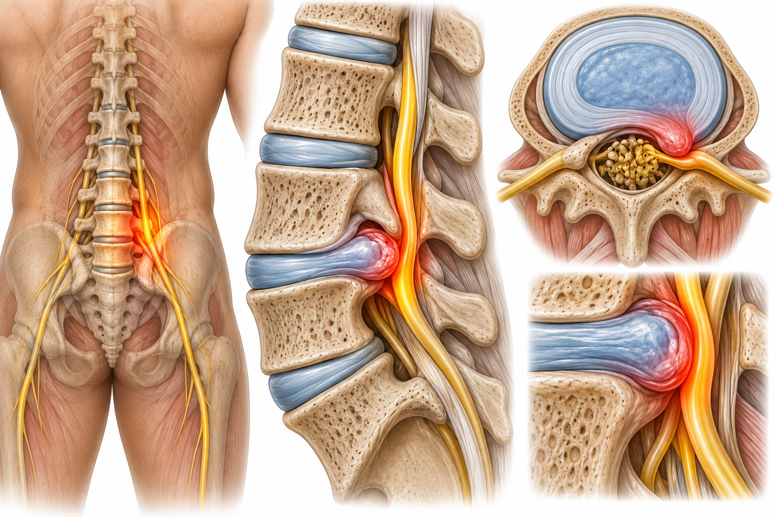
The intervertebral disc is a fibrocartilaginous structure between adjacent vertebrae, composed of a tough outer ring — the annulus fibrosus — and a gel-like inner core — the nucleus pulposus. A disc bulge occurs when the nucleus pushes outward, deforming the disc without fully breaching the annulus. A herniation — also called a prolapse or slipped disc — occurs when the nucleus ruptures through the annular fibres and contacts the adjacent neural tissue.
The critical clinical point is that disc findings on MRI are extremely common in people without any symptoms. Studies show that the majority of people over 40 have disc bulges visible on imaging — most of which are completely asymptomatic. A disc becomes clinically significant only when it is compressing the correct nerve root and producing symptoms that match the anatomical territory of that root.
ROOT METHOD's approach to disc pathology is to confirm clinical significance before any treatment is started. The combination of symptom distribution, nerve tension testing, dermatomal mapping, and reflex assessment establishes whether the disc identified on imaging is genuinely responsible for your specific symptoms — or whether the real pain source lies elsewhere.
Clinical Presentation
Common Symptoms of Disc Herniation
Disc herniation symptoms vary depending on whether the cervical or lumbar spine is affected, and which nerve root is compressed. Each symptom carries specific diagnostic significance.
Sharp Spinal Pain
Acute pain localised to the affected disc level, characteristically worse with forward flexion, prolonged sitting, and Valsalva manoeuvres. The pain may be severe at onset and is often accompanied by protective muscle spasm and an antalgic posture.
Radiating Arm or Leg Pain
Cervical disc herniation produces arm pain in the distribution of the affected cervical nerve root. Lumbar herniation produces leg pain following the sciatic or femoral nerve territory. The pattern of radiation is the most powerful indicator of which disc level is involved.
Neurological Deficit
Numbness or altered sensation in a specific dermatome — a precise band of skin supplied by the compressed nerve root. Dermatomal sensory loss is one of the most diagnostically precise signs in spinal assessment, reliably identifying the affected level.
Muscle Weakness
Weakness in the myotomal distribution of the compressed nerve root — for example, wrist extension weakness with C6 compression, or big toe extension weakness with L5 involvement. Myotomal weakness confirms functional nerve compromise and influences the urgency of clinical management.
Pain with Valsalva
A characteristic and highly specific feature — symptoms significantly worsen with coughing, sneezing, or straining. These manoeuvres increase intrathecal pressure and load the disc, transiently increasing nerve root compression and producing a sharp exacerbation of pain.
Antalgic Posture
Patients frequently adopt a lateral lean — away from the side of the herniation — as a subconscious strategy to reduce nerve root contact. This antalgic lean is a visible clinical sign that, combined with movement testing, helps confirm disc pathology and identify which side the herniation is on.
Clinical Aetiology
Common Causes of Disc Herniation
Disc herniation results from a combination of structural vulnerability and mechanical load. Understanding the mechanism guides both the diagnosis and the management approach.
Annular Tear
A radial tear through the concentric layers of the annulus fibrosus allows the pressurised nucleus pulposus to track outward. This is the fundamental mechanical event in disc herniation — and can occur acutely with a single load or gradually through cumulative annular damage.
Age-Related Degeneration
With age, the nucleus pulposus loses water content and the annulus becomes more brittle and less elastic. This degeneration reduces the disc's ability to withstand compressive and rotational loads — making herniation more likely even without significant trauma.
Acute Compressive Load
Heavy lifting combined with rotational stress — particularly bending and twisting simultaneously — applies peak compressive and shear force to the posterior annulus. This is the classic mechanism of acute disc herniation, often occurring at a clearly identified moment during a specific activity.
Repetitive Flexion
Sustained or repeated forward bending — common in occupations requiring prolonged sitting or manual handling — applies cyclical posterior pressure to the disc nucleus. Over time, this can drive the nucleus progressively toward the posterior annulus, creating a gradual herniation without a single identifiable traumatic event.
Disc Height Loss
As disc height is lost through degeneration, adjacent facet joints become overloaded — carrying forces that were formerly distributed through the disc. This facet overloading compounds disc degeneration and contributes to foraminal narrowing, further increasing the risk of nerve root compression even without frank herniation.
Inflammatory Response
The nucleus pulposus contains inflammatory mediators including phospholipase A2 and cytokines. When it contacts a nerve root, it produces a chemical radiculitis — inflammation of the nerve independent of mechanical compression. This is why some patients have severe symptoms from small herniations that appear to barely touch the nerve on imaging.
Clinical Guidance
When to Seek Professional Assessment
Disc pathology ranges from self-limiting to surgical. Knowing when clinical assessment is needed — and when it is urgent — is essential.
Book a Clinical Assessment If…
- Radiating arm or leg pain has persisted for more than 6 weeks
- You have neurological symptoms — numbness, tingling, or weakness in the limb
- An MRI has shown a disc herniation and you have not received a clinical interpretation of what it means for your specific symptoms
- Pain significantly worsens with coughing, sneezing, or sitting
- Prior physiotherapy or injections have not produced lasting improvement
- You are considering surgery and want an independent clinical assessment first
Seek Emergency Care Immediately If…
- You develop sudden loss of bladder or bowel control — cauda equina syndrome requires emergency surgical decompression
- Progressive bilateral leg weakness or numbness develops over hours
- Saddle anaesthesia — numbness in the perineum, inner thighs, or genitals
- Rapid deterioration of motor function in the arm or leg — do not wait for a scheduled appointment

The ROOT METHOD Difference
How ROOT METHOD Approaches Disc Herniation
The single most common clinical error with disc pathology is treating the disc seen on imaging without confirming that it is actually responsible for the patient's symptoms. ROOT METHOD addresses this by performing the clinical investigation first — mapping the symptom pattern to the anatomy before any imaging is interpreted.
Every assessment identifies: the specific disc level involved, the nerve root affected, the degree of functional compromise, and whether conservative management is appropriate or surgical referral is indicated.
- Nerve tension testing — SLR, slump test, upper limb tension tests — to confirm nerve root involvement
- Dermatomal sensory mapping — precisely identifying the affected nerve root by its territory
- Myotomal strength testing — grading motor compromise and monitoring for deterioration
- Directional movement testing — identifying which movements centralise pain (a key guide to management)
- Surgical threshold assessment — clear criteria for when conservative treatment is appropriate and when referral is needed
Your First Visit
What to Expect During Your Disc Assessment
A ROOT METHOD disc herniation assessment is a 90-minute clinical appointment. Here is what happens at each stage.
-
Before You Arrive
Pre-Visit Intake Form
Complete the ROOT METHOD 8-step intake form, including details of your pain onset, symptom distribution in the arm or leg, what aggravates or relieves your symptoms, and any prior imaging or treatment. Bring any existing MRI reports — Dr. Umer will incorporate them into the clinical reasoning process.
-
Stage 1
Movement and Directional Assessment
Spinal movements are assessed in all planes — identifying which directions reproduce, worsen, or relieve your radiating symptoms. Centralisation of pain with extension or lateral flexion is a powerful diagnostic and prognostic finding that guides both the diagnosis and the treatment approach.
-
Stage 2
Neurological and Structural Examination
Full nerve tension testing, dermatomal sensory assessment, deep tendon reflexes, and myotomal strength grading are performed. Findings are mapped to specific spinal levels, producing a precise clinical picture of which disc is involved and how significantly the nerve root is compromised.
-
Stage 3
Diagnosis and Clinical Pathway
Dr. Umer explains the clinical findings, correlates them with your imaging if available, and presents a clear management pathway — whether conservative rehabilitation, a supervised directional exercise programme, or a surgical referral with documented clinical justification.

Common Questions
Questions About Disc Herniation and ROOT METHOD
Is a disc bulge the same as a herniation?
Not precisely, though both terms describe disc protrusion. A disc bulge is a broad-based, symmetrical outward expansion of the disc beyond its vertebral margins — the annulus is intact but deformed. A herniation involves a more focal protrusion where the nucleus has breached or significantly strained the annular fibres. A frank herniation — also called a prolapse or extrusion — occurs when nucleus material fully ruptures through the annulus.
In clinical practice, the distinction matters less than whether the disc is causing nerve root compression. A modest focal herniation that contacts a nerve root is more clinically significant than a broad bulge that does not.
Will a disc herniation heal on its own?
Yes, in many cases. The body's immune system recognises herniated nucleus pulposus as foreign material and progressively resorbs it. Studies show that approximately 60–80% of disc herniations show measurable reduction on MRI within one year without surgical intervention. Larger herniations — particularly sequestered fragments that have fully separated from the disc — often resorb more completely than smaller contained herniations.
Conservative management supports this natural recovery by reducing nerve root inflammation, maintaining spinal mobility, and identifying directional exercises that reduce symptom severity during the healing process.
Does MRI always show the problem?
Not reliably, in either direction. MRI over-detects — showing disc changes in people without pain — and under-detects clinically relevant pathology in some presentations, particularly when the herniation is small or at an unusual location. Crucially, the severity of findings on MRI does not correlate predictably with the severity of clinical symptoms.
This is why clinical examination is the foundation of ROOT METHOD's approach. MRI is an important tool — but it answers the question of anatomy, not the question of which anatomy is causing your specific symptoms. Only a thorough clinical examination can answer that.
Do I need surgery for a disc herniation?
Most disc herniations do not require surgery. The indications for surgical intervention are specific: progressive neurological deficit (worsening weakness or numbness), failure to improve after an adequate trial of appropriately targeted conservative treatment (typically 6–12 weeks), or cauda equina syndrome, which requires emergency surgery.
ROOT METHOD's assessment establishes from the outset whether surgery is indicated based on clinical criteria — not on imaging findings or symptom duration alone. Patients who meet criteria are referred with documented clinical justification. Those who do not are given a conservative protocol with clear milestones.
How do I know which level my disc problem is at?
The clinical examination provides a strong indication of the affected level through dermatomal mapping, myotomal strength testing, and reflex assessment. L4 root compression produces medial calf sensory change and reduced knee reflex. L5 produces dorsal foot and big toe changes with weakness of big toe extension. S1 produces lateral foot and heel sensory change with reduced ankle reflex.
In the cervical spine, each root level produces a characteristic arm and hand sensory pattern that an experienced clinician can map reliably. Imaging then confirms the clinical suspicion and localises the pathology precisely.
How long is recovery from disc herniation?
Recovery timeline depends on the degree of nerve root compression, the presence of neurological deficit, and whether treatment is correctly targeted. Acute disc herniation with radiculopathy typically improves significantly within 6–12 weeks of appropriately directed management. Neurological signs — numbness and weakness — often resolve more slowly than pain, sometimes taking 3–6 months to fully normalise.
Patients whose treatment has been targeted at an incorrect diagnosis may remain symptomatic far longer. Establishing the correct clinical picture early, and targeting treatment accordingly, is the most important factor in achieving a complete and durable recovery.
Your Disc Pain Has a Level. We Identify It.
A disc finding on MRI is not a diagnosis. ROOT METHOD determines which disc is clinically significant, which nerve root is involved, and whether conservative management will resolve your symptoms — before any treatment decision is made.